This section highlights selected projects involving 3D modeling, animation, and interactive visualization. These works translate complex scientific and medical subjects into accessible visual tools for research communication, education, and public engagement.
Animals in Research - Educational Visualization
(University of Dundee & NC3RS)
This project was developed for the Animals in Research program at the University of Dundee as part of a public engagement initiative aimed at improving transparency and accessibility around the role of animals in biomedical research. The project involved redesigning an existing informational pamphlet into an illustrated flipbook format and creating a short educational animation intended for public audiences.
Using illustrated rats and mice, the materials explain how these animals contribute to scientific research while presenting the information in a clear, approachable visual style. The updated visuals were designed to make complex and often sensitive topics easier to understand, supporting the university’s goal of fostering informed public discussion around animal research.
The project involved redesigning an existing informational pamphlet into an illustrated flipbook format and producing a new animated video to support public engagement and accessibility.
(Original Flipbook)
(Redesigned Flipbook)
Research & Concept Development
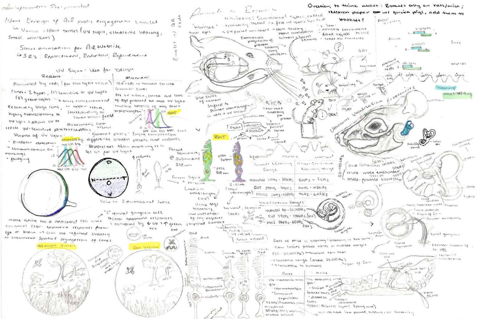
Early development for this project focused on understanding the biological mechanisms behind key sensory systems in laboratory rodents. Preliminary studies included anatomical drawings exploring visual processing (eye structure and the function of rods and cones), auditory anatomy within the inner ear, and the tactile system of whiskers, tracing how vibrations travel through follicles and neural pathways to the brain and more. These concept sketches informed the final visual approach by grounding the illustrations and animation in accurate biological function while simplifying complex processes for clear public communication.
Animation Stills
Arterial Thoracic Outlet Syndrome – Medical Visualization
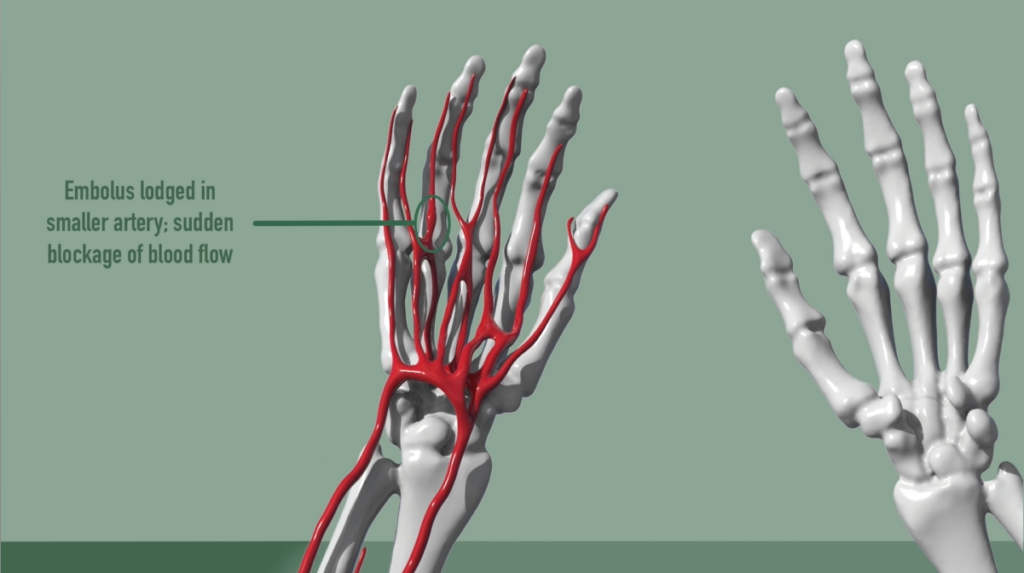
This project explores the anatomy and pathology of arterial thoracic outlet syndrome (ATOS) through a combination of anatomical illustration and animation. The work includes a detailed educational poster and an animated video designed to communicate the anatomical structures involved and the mechanisms through which vascular compression can occur in the thoracic outlet region.
By combining clear anatomical visualization with motion-based explanation, the project aims to make a complex vascular condition easier to understand for students and general audiences. The materials demonstrate how medical visualization can support education by translating complex anatomical relationships into accessible visual formats.
Educational Animation
Condition Overview
Arterial thoracic outlet syndrome (ATOS) is a rare vascular condition that occurs when the subclavian artery becomes compressed as it passes through the thoracic outlet, the narrow space between the clavicle and the first rib. This compression may be caused by anatomical variations, muscular structures such as the scalene muscles, or abnormal bone formations including cervical ribs.
Anatomical Poster
When the artery is compressed, normal blood flow to the arm can be disrupted, potentially leading to symptoms such as pain, numbness, cold sensitivity, weakness, or reduced circulation in the affected limb. In severe cases, arterial damage or blood clots may develop.
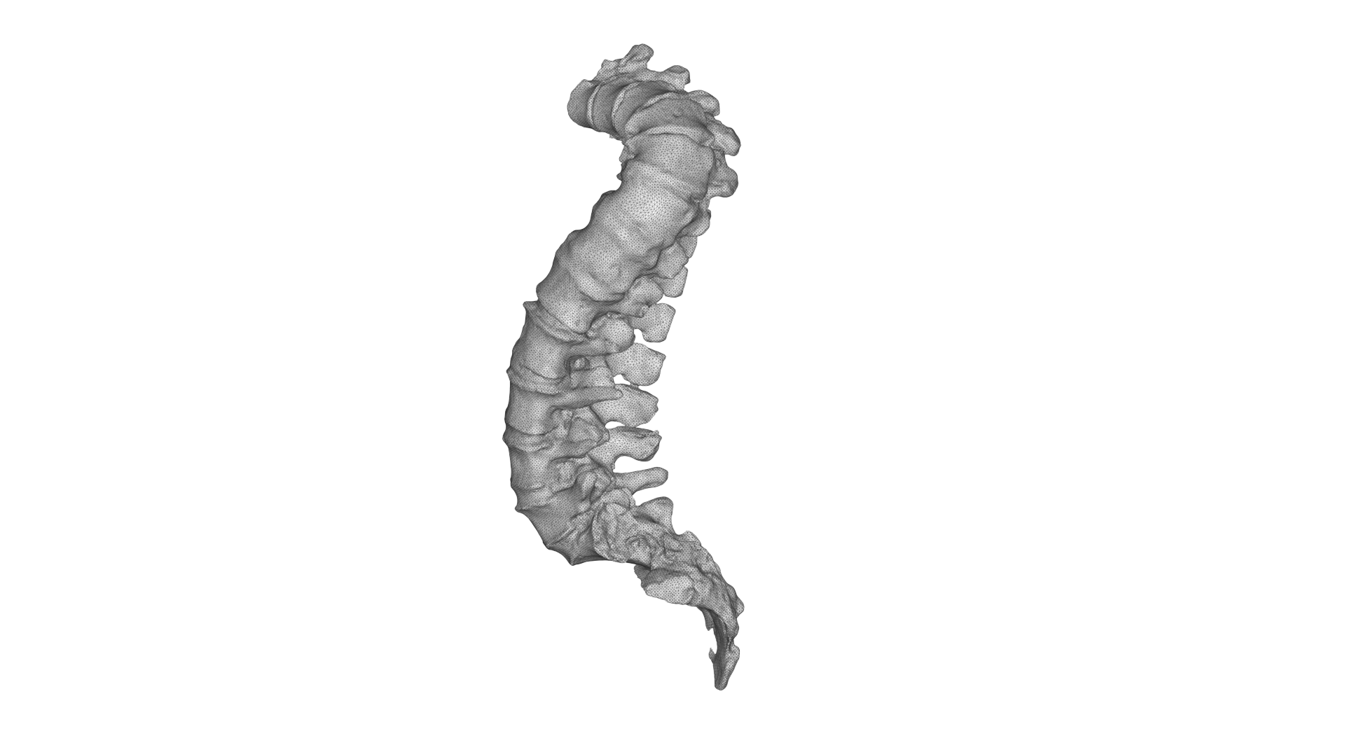
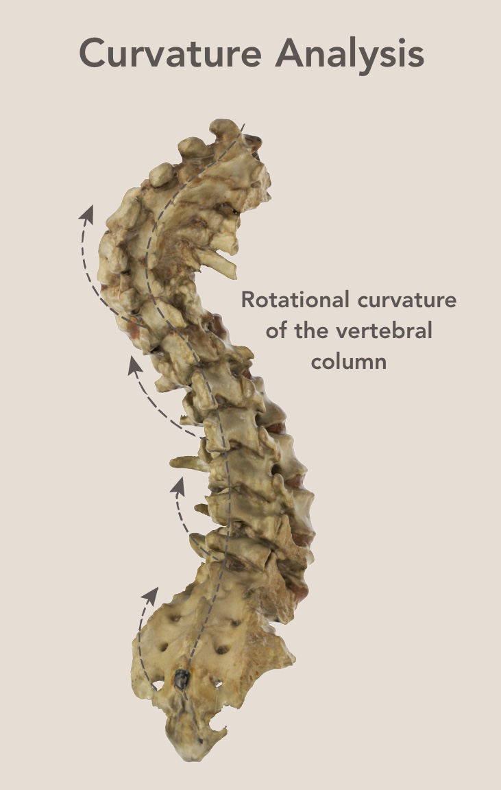
This project involved the digital scanning and presentation of a severely twisted spinal column specimen for educational reference at CAHID (Centre for Anatomy and Human Identification) in Scotland. Using Artec scanning technology, the specimen was captured and converted into an interactive 3D model that allows viewers to examine the structure from multiple perspectives.
The model was labeled to highlight key anatomical landmarks and structural deviations, creating a visual reference tool for studying spinal deformities. By presenting the specimen in an interactive digital format, the project demonstrates how 3D scanning can preserve and extend access to anatomical material for educational and research contexts.
Model Processing & Cleanup
Following the scan capture, the model was processed to refine and prepare the geometry for visualization. This included aligning scan data, removing artifacts, filling gaps, and optimizing the mesh to create a clean and readable anatomical model. The final processed model was then prepared for interactive viewing and anatomical labeling.
Artec 3D Scanning
The spinal column specimen was digitized using Artec 3D scanning technology, capturing high-resolution surface geometry of the vertebrae and surrounding structures. Multiple scan passes were collected and aligned to accurately document the complex curvature and rotation of the spine, producing a detailed digital representation of the specimen.
Anatomical Labelling & Educational Visualization
Pangolin Anatomy & Adaptation Visualization
Interactive Biological Visualization Study
(D’Arcy Thompson Zoology Museum)
Defensive Behavior
Pangolins are the only mammals covered in protective keratin scales and possess one of the longest tongues relative to body size in the animal kingdom.
This project explores the distinctive anatomy and behavioral adaptations of pangolins through a combination of 3D modeling, anatomical visualization, and animation. Pangolins are highly specialized mammals known for their protective keratin scales, powerful digging forelimbs, and unique feeding adaptations that allow them to consume ants and termites.
The study includes comparative 3D models demonstrating both quadrupedal locomotion and an upright defensive posture, highlighting how body structure supports different behaviors. Additional anatomical visualizations examine skeletal features of the forelimbs, skull, and spine, as well as the pangolin’s exceptionally elongated tongue, which extends deep into the body cavity as part of its specialized feeding mechanism.
A short animated sequence further demonstrates the pangolin’s characteristic defensive behavior—curling into a tight ball to shield its vulnerable underside. Together, these visualizations aim to present the biological adaptations of pangolins in a clear and engaging format, combining accurate anatomical study with accessible scientific communication.
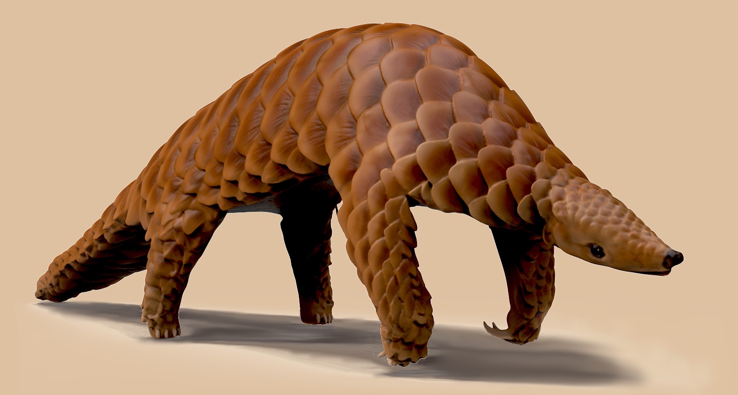
Locomotion & Posture
Pangolins primarily move on four limbs but are capable of rearing upright when feeding or assessing threats by using their tail as both a counterweight and additional limb.
Upright/ Defensive Pose
Quadrupedal Pose
Skeletal Adaptations for Digging and Feeding
Comparative Posture
Comparative poses demonstrating pangolin locomotion and upright defensive posture.
3D Models, Anatomical Section Studies, and Educational Animation
This series explores normal kidney anatomy and the structural changes associated with polycystic kidney disease (PKD). Interactive models and animations illustrate how the kidney filters blood, produces urine, and how cyst formation disrupts normal function.
In polycystic kidney disease, fluid-filled cysts progressively enlarge and distort the normal structure of the organ.
Internal Structure
Sagittal sections reveal the internal organization of the renal cortex, medulla, and collecting system. In polycystic kidney disease, expanding cysts progressively replace functional tissue and compress surrounding structures.
Whole Kidney →Fade to Sagittal Cut
Mobile Viewers, click to view
Whole Kidney → Fade to Sagittal Cut
Mobile Viewers, click to view
Kidney Anatomy
The kidney filters blood through millions of microscopic filtration units known as nephrons. Blood plasma enters the nephron through the glomerulus, where filtration begins before fluid travels through the renal tubules to form urine.
Urine Formation
Urine formation begins in the nephron, where blood plasma is filtered in Bowman’s capsule before passing through a series of specialized tubules that regulate water, salts, and waste products.
Animation Still
Polycystic Kidney Disease
Polycystic kidney disease is a genetic disorder characterized by the progressive development of fluid-filled cysts throughout the kidneys. Over time, these cysts enlarge and disrupt normal kidney function, potentially leading to kidney failure.